
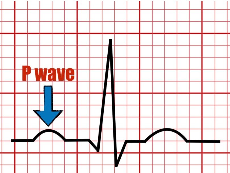
The P Wave

In this article, you will learn about the meaning and characteristics of the P wave. I will also mention some disease states and arrhythmias that may lead to an abnormal or absent P wave.
P wave definition
The P wave represents atrial depolarization which stimulates contraction of the atriums. It can be seen as the first deflection in the P-QRS-T complex. Depolarization wave starts from the sinoatrial node (SA) and travels along the interatrial septum and reaches the left atrium. Given that the SA node is located in the right atrium, this chamber is the first to begin to depolarize.
The P wave is divided into two components representing depolarization of the right and left atrium.

P wave on lead II

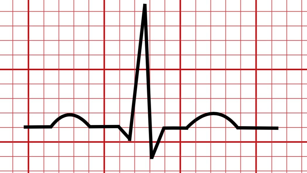
P wave on lead V1
P wave axis
The normal P wave axis is 60 ° but may vary between 0 to +75°.
Upright in lead II and usually in leads I, aVL, aVF.
Inverted in lead aVr.
Biphasic in lead V1 and V2.

Normal P wave axis 0 - +75°
P wave size and duration

Normal P wave parameters
The normal P wave duration: 0.08 - 0.11 second

Abnormal P wave
Right atrial enlargement
Positive deflection in lead v1 or v2 > 1.5 mm
Tall upright P wave >2.5 mm in lead II
Left atrial enlargement
Negative deflection in V1 > 1 mm wide (duration>0.04s) and > 1 mm deep (amplitude).
Duration of the P wave > 120 ms or more with a widely notched P wave, 40 ms or more in lead II.
Several disease states that may alter the P wave amplitude and duration :
Hypertension
Ischemic Heart Disease
Pulmonary hypertension
Valvular disease
COPD
Congenital heart disease
Sinus P wave may be absent in several arrhythmias, including:
Atrial flutter
Atrial fibrillation
Wandering Pacemaker
Multifocal atrial tachycardia
Atrial tachycardia
Junctional rhythm
Key Takeaways
The P wave represents atrial depolarization leading to atrial contraction.
Both right and left atrial components are embedded in the P wave.
The normal axis is between 0 - +75°.
The P wave is normally upright in leads I, II, and III, biphasic in leads v1 and v2, and inverted in lead aVr.
Any deviation from the normal amplitude and duration of the P wave may suggest an atrial abnormality.
Several disease states and arrhythmias may alter the normal P wave parameters or even lead to its absence.
References
1. Hancock EW, Deal BJ, Mirvis DM, et al. AHA/ACCF/HRS Recommendations for the Standardization and Interpretation of the Electrocardiogram part V. J Am Coll Cardiol. 2009;53:992-1002.
2. Surawicz, B., Knilans, T. K., & Chou, T.-C. (2008). Chou's electrocardiography in clinical practice: Adult and pediatric. Philadelphia, PA: Saunders/Elsevier.
3. O’Keefe J, Hammill S, Freed M. (2016). The Complete Guide of ECGs (4th ed.). Jones & Bartlett Publishers.
Please send your feedback !
In this blog, we are going to explain the meaning of the U wave in the electrocardiogram and provide examples of normal and pathologic causes of its appearance on the ECG.