
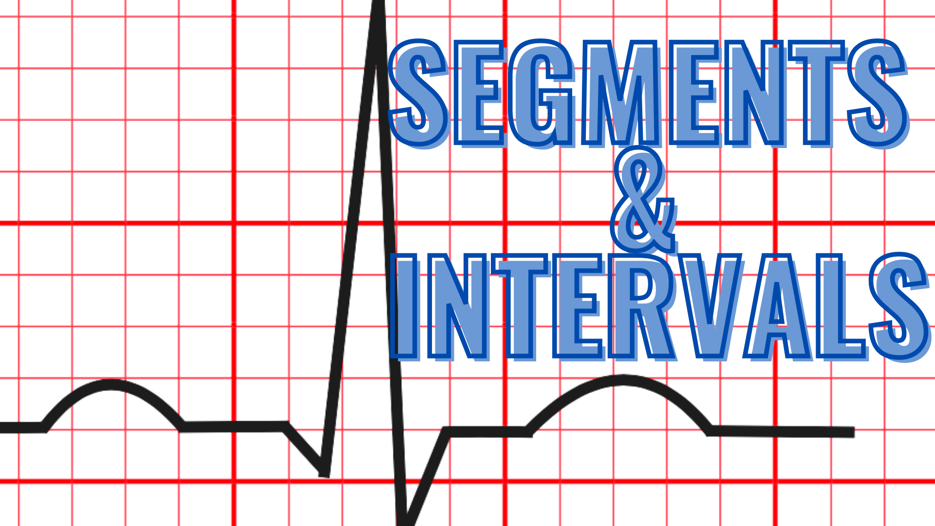
ECG Segments and Intervals

In this blog, we are going to explain the meaning of the segments and intervals on the ECG and discuss each of them in detail. This information will help the ECG interpreter to correlate each segments and intervals to its correspondent cardiac events and learn some pathologic conditions that may alter their normalcy.
What are the segments of an ECG?

Segments of the ECG
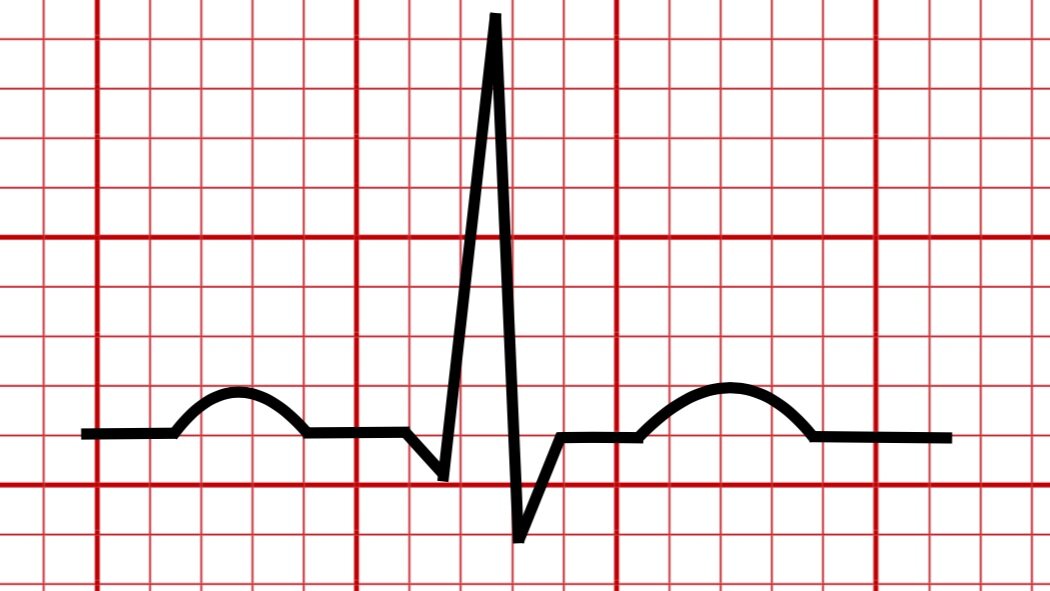
The segments on an ECG are nearly isoelectric lines that connect each wave.
There are three different segments on the ECG
The PR segment
The ST segment
The TP segment
The PR segment

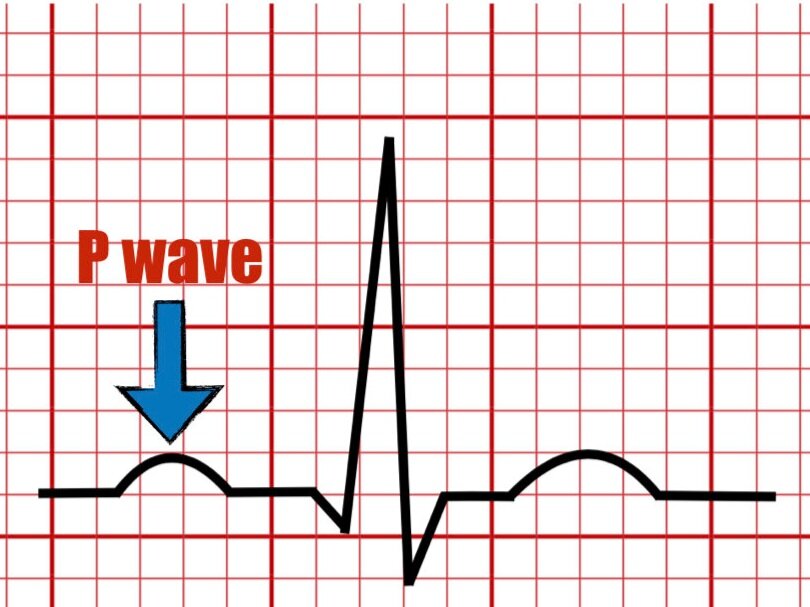
The PR segment starts at the end of the P wave and ends at the beginning of the QRS complex.
The PR segment represents the end of atrial depolarization, and the beginning of ventricular depolarization, forming a temporal bridge between atrial activation and ventricular activation.
Inside the PR segment several events are embedded including atrial repolarization, slow conduction through the AV node, and rapid conduction along with the ventricular conduction system.
Atrial injury or pericarditis may lead to changes in the isoelectric line of the PR segement.
The ST segment

The ST segment is the isoelectric line starting at the end of the QRS complex (J point) to the beginning of the T wave.
The ST segment represents a temporal bridge between ventricular depolarization and ventricular repolarization.
The normal ST segment configuration can be altered in early repolarization, subendocardial ischemia, ST-elevation myocardial infarction, and pericarditis.
The TP Segment

The TP segment is the isoelectric line that begins at the end of the T wave and ends at the start of the P wave.
The TP segment can be used as a reference for evaluation or pathologic conditions such as pericarditis or atrial injury.
The slope of the TP segment can be altered during phase 1 of acute pericarditis (Spodick’s sign).
What are the intervals on an ECG?

Intervals of the ECG
The intervals on the ECG refers to the duration of a cardiac event reflected on an ECG tracing.
Intervals in the ECG include at least one segment and wave.
Several pathologic states may alter the normal duration of intervals.
The intervals on the ECG are
PR interval
RR interval
PP interval
QT interval
The PR Interval

The PR interval or PQ interval begins at the onset of the P wave (atrial depolarization) and ends a the beginning of the QRS complex (ventricular depolarization).
It reflects the atrial contraction during late diastole and slow AV conduction.
Prolongation of this interval can be seen on first degree AV block.
Short PR intervals have also been seen on WPW.
Normal duration 120 milliseconds (ms) - 200 ms.
The RR interval

The RR interval is the time interval between two successive R waves or two systolic events.
Can be used for reference when calculating the corrected QT interval.
The RR interval duration may vary inversely with heart rate, i.e., faster heart rate leads to shorter RR intervals and vice versa.
The RR interval is useful for evaluating the heart rate variability which reflects neuroautonomic control of the heart beat.
The PP Interval
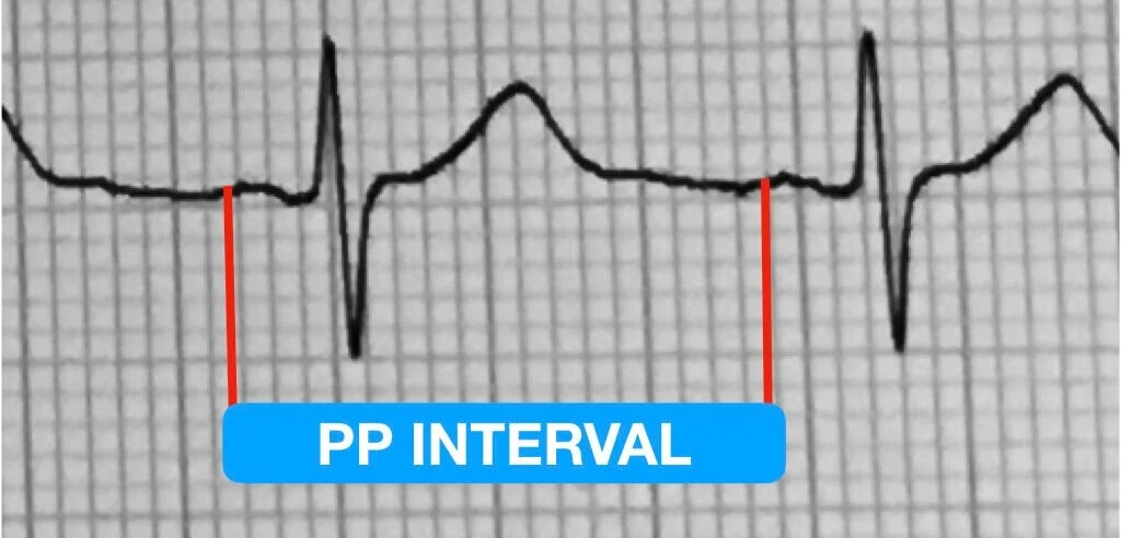
The PP interval is the interval between two successive P waves (atrial contraction).
The PP interval can vary with respiration (respiratory sinus arrhythmia).
The PP interval should be identical to the RR interval in sinus rhythm.
The QT Interval

The QT interval starts at the beginning of the QRS complex to the end of the T wave.
The QT interval represents the duration of ventricular electrical systole (ventricular depolarization and repolarization).
The duration of the QT interval should be less than half the RR interval.
The normal QT interval varies with heart rate ,i.e., fasters heart rates leads to shorter QT intervals..
Within a heart rate of 60 beats-per-minute (bpm) - 100 bpm the normal duration of the normal QT interval is less than 450 ms for men and 460 ms for women.
The QT interval is prolonged when the corrected QT interval (QTc) duration is more than 440 ms in men and 460 ms in women.
Short QT interval is considered when QTc is less than 390 ms in both men and women.
The QT interval can be prolonged genetically or with certain medications or electrolyte imbalances.
References
Rautaharju PM, Surawicz B, MD, Gettes LS. Recommendations for the standardization and interpretation of the electrocardiogram: part IV: the ST-Segment, T and U Waves, and the QT Interval. J Am Coll Cardiol. 2009:53:982–91.
Surawicz, B., Knilans, T. K., & Chou, T.-C. (2008). Chou's electrocardiography in clinical practice: Adult and pediatric. Philadelphia, PA: Saunders/Elsevier.
Zipes, D. P., Libby, P., Bonow, R. O., Mann, D. L., Tomaselli, G.F., &Braunwald, E. (2019). Braunwald's heart disease: A textbook of cardiovascular medicine (Eleventh edition.). Philadelphia, PA: Elsevier/Saunders.

In this blog, we are going to explain the meaning of the U wave in the electrocardiogram and provide examples of normal and pathologic causes of its appearance on the ECG.