
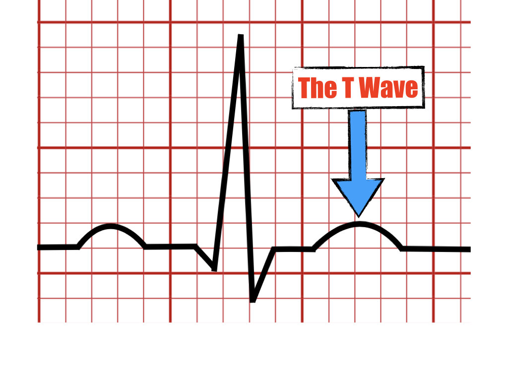
The U wave

The U wave is found after the T wave.
Begins from baseline after the end of T wave.
It was first described by Einthoven in the year (1903).
Its amplitude is 0.33mV or 11% of the T wave and has the same polarity as the T wave.
The U wave represents the end of repolarization and is best seen in the precordial leads V2-V3.
The U wave is rarely seen in 50-75% of cases.
It is heart rate dependent, 90% of cases with HR below 65 bpm and rarely found with HR above 95 bpm.

Arrows depict U waves on leads V3 - V4
Origin of the U wave
Several theories have been postulated regarding the origin of the U wave but it is still controversial. Some theories include
Delayed afterdepolarization of the His-Purkinje system
Repolarization of the papillary muscle
Prolonged repolarization in the mid myocardium cells (M cells)
Prominent U wave
Can be seen on several instances including
Hypokalemia
Class Ia and III antiarrhythmics
LQTS
Bradycardia
Hypothermia
Left ventricular hypertrophy
Normal variant
Negative U waves
Negative U waves have been associated with heart disease specifically
Myocardial ischemia
Myocardial infarction
Ventricular hypertrophy
Valvular disease
Hypertension.
References
AHA/ACCF/HRS Recommendations for the Standardization and Interpretation
of the Electrocardiogram Part IV: The ST Segment, T and U Waves, and the QT Interval
Cardiovasc Metab Sci. 2021; 32(4): 197 - 205
Surawicz B. U wave: Facts, hypotheses, misconceptions, and misnomers. J Cardiovasc Electrophysiol. 1998; 9 (10): 1117-1128.
Duque-González L, Gaviria-Aguilar MC, Lopera-Mejía L, Duque-Ramírez M. The U wave: an ignored wave flled with
information. Cardiovasc Metab Sci. 2021; 32 (4): 197-205.
Carrillo-Esper R, et al. The U wave in the electrocardiogram. More than an academic curiosity. Rev Invest Med Sur Mex, 2015; 22 (1): 27-29
In this blog, we are going to explain the meaning of the U wave in the electrocardiogram and provide examples of normal and pathologic causes of its appearance on the ECG.